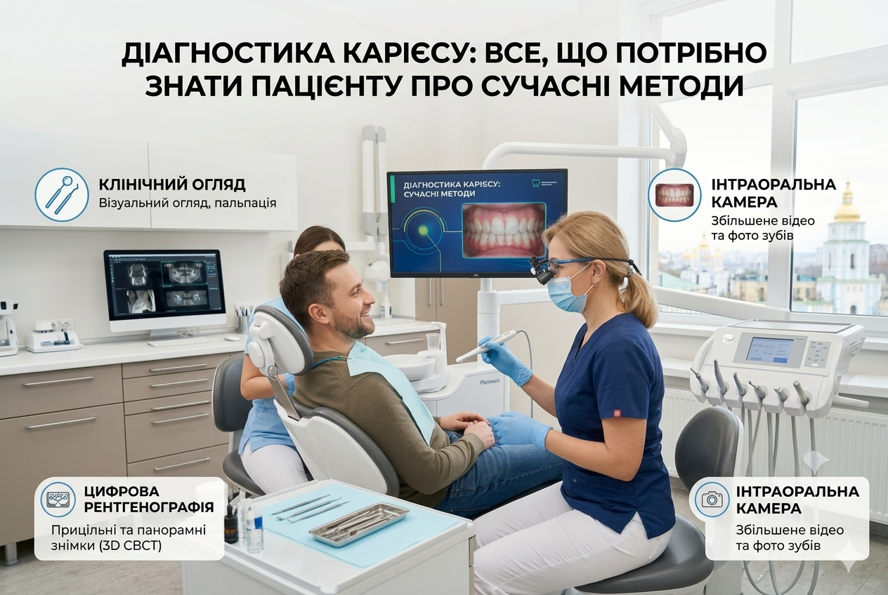
Caries Diagnosis: Everything a Patient Needs to Know About Modern Methods
Caries is a treacherous enemy. It does not appear overnight and does not always announce itself with sharp pain. Patients often follow the iron rule: “If it doesn’t hurt, don’t touch it.” However, in the case of teeth, this rule only works to increase the cost of your future treatment. When pain appears, it is no longer about a simple filling, but about root canal treatment (pulpitis).
Modern dentistry is focused on micro-invasiveness: detecting the problem at an early stage and preserving as much healthy tooth tissue as possible.
Why is a simple mirror examination no longer enough?
Previously, diagnosis looked simple: the doctor would look at the tooth with a mirror, poke it with a sharp probe, and if it got stuck somewhere, they would deliver a verdict. Today, this method is considered outdated and even dangerous, as the probe can damage weakened enamel that might otherwise have recovered (remineralized).
Furthermore, caries loves to hide in hard-to-reach places:
- In fissures — natural depressions on the chewing surfaces (while the enamel on the surface may appear completely intact).
- Between teeth (contact caries) — the most insidious type, which is often noticed only when a tooth wall simply breaks off while eating.
- Under old fillings and crowns (secondary caries).
Top diagnostic methods in a modern clinic
Today, a dentist is somewhat of a detective with a bunch of gadgets. Here is what helps doctors see the hidden:
- Digital radiography and 3D tomography (CT)
This is the “gold standard.” On a computer screen, a doctor can clearly discern the internal structure of the tooth. An X-ray will show caries between teeth at a stage when it is not yet visible, while a CT scan allows for assessing the depth of the lesion and the condition of the tissues around the root. - Dental microscope and loupes
Optics increase the working field by 10–20 times. The doctor uses special binocular glasses. This allows them to notice micro-cracks in the enamel, detect initial demineralization (a “white spot”), and check the fit of an old filling to see if there are micro-gaps where bacteria can accumulate. - Laser fluorescence (DIAGNOdent)
The device illuminates tooth tissues with a laser. Healthy enamel and damaged enamel reflect light waves of different lengths. The device analyzes this and provides a digital index: the doctor knows exactly whether it is just pigmentation (which does not need to be touched) or if it is already caries. - Caries markers
These are special safe organic dyes. The doctor applies them to the teeth during treatment or an examination. The marker stains only the dentin destroyed by bacteria and is easily washed off healthy tissues. This helps the dentist not remove excess tissue during drilling.
Warning signs: when to run to the doctor?
Do not wait for a scheduled visit if you notice at least one of these symptoms:
- Reaction to irritants: short-term pain or a “tingling” sensation from cold, hot, sweet, or acidic foods that disappears immediately after the source is removed.
- Problems with floss: if during the cleaning of interdental spaces, the dental floss constantly gets caught on something, breaks, or shreds.
- Visual changes: the appearance of chalky (matte-white) or dark spots on the enamel.
- Bad breath: an unpleasant odor from the mouth that does not disappear even after thorough brushing.
Important: Modern diagnostics is a fast, painless, and highly accurate process. A preventive check-up every six months using optical and digital methods will allow you to “catch” caries at a stage when its treatment requires a minimum of time and money.